
Medically reviewed by Dr. Mac Maxwell, DACVS — board-certified veterinary surgeon. Last reviewed: June 2026
Quick Facts: Medial Patellar Luxation (MPL)
- What it is: A common knee condition in dogs where the kneecap (patella) slips inward, medially, out of its normal groove.
- Most affected: Small and toy breeds, including Boston Terriers, Yorkshire Terriers, Pomeranians, Miniature Poodles, and Chihuahuas.
- Grades I to IV: From Grade I (luxates only with manual pressure, returns on its own) to Grade IV (permanently out of place).
- Common signs: Skipping or hopping on a back leg, intermittent hind-leg lameness, and reluctance to jump or run.
- Treatment: Mild cases (Grade I, some Grade II) are managed without surgery; Grade III, Grade IV, and symptomatic Grade II usually need surgical correction.
- Recovery: Most dogs need about 8 to 12 weeks of restricted activity and return to normal activity in roughly three months.
- Outcomes: Reported owner satisfaction after surgical correction is commonly over 90%.
Medial patellar luxation (MPL) is one of the most common orthopedic conditions we see in dogs, particularly small and toy breeds. This guide explains what MPL is, how it’s graded, the signs to watch for, and when surgical correction is the right step. At Apex Veterinary Surgery, our board-certified (DACVS) surgeons evaluate and treat MPL for pets across Memphis, Nashville, and surrounding areas of the Midsouth.
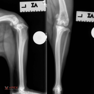
A lateral and AP radiograph view of a dog’s stifle/knee. The patella (kneecap) normally rides in the trochlear groove at the end of the femur; with medial patellar luxation, it slips inward out of that groove. The front-to-back view shows the kneecap relative to the groove and is where a medial (inward) luxation is most clearly seen.
Understanding Medial Patellar Luxation
The patella (kneecap) is a small bone that sits within the tendon of the quadriceps muscles at the front of the thigh. Normally, it rides in a groove (the trochlear groove) at the end of the femur as the knee bends and straightens. The patellar tendon anchors to the tibial crest, a bony ridge just below the knee. Together, the quadriceps muscle, patella, and patellar tendon form the “extensor mechanism,” which is normally well aligned. Patellar luxation is the dislocation of the kneecap out of its groove when the knee flexes.
In a medial luxation, the kneecap slips inward, toward the dog’s body. MPL is classified into four grades by severity:
- Grade I: The patella can be manually pushed out of the groove but returns to position on its own when released.
- Grade II: The patella luxates occasionally on its own, causing intermittent lameness, and can be replaced by manipulation. In Grade II’s the patella spends most of the time in the normal position.
- Grade III: The patella sits out of the groove most of the time, causing persistent lameness, but can still be manually replaced. In Grade III, the patella spends most of the time in the luxated or medial position.
- Grade IV: The patella is permanently luxated and cannot be manually returned to the groove, leading to significant discomfort and limb deformity.
Why Patellar Luxation Happens
MPL usually results from a combination of genetic and developmental factors, and less often from trauma. Predisposed breeds include Boston Terriers, Yorkshire Terriers, Pomeranians, Miniature Poodles, and Chihuahuas, among others. The underlying cause is typically a malalignment in the structures of the limb, such as the femur, tibia, or the soft tissues that support the knee, which pulls the patella out of the trochlear groove.
Signs Your Dog May Have an MPL
Symptoms vary widely in severity. In mild cases, MPL may be an incidental finding during a routine exam. Signs to watch for include:
- Limping or lameness in one or both hind legs, often intermittent at first and becoming more frequent as the condition progresses
- An abnormal gait, such as skipping or hopping, sometimes carrying the leg up for a few steps before setting it back down
- Reluctance to jump or run
- A “bow-legged” stance, especially in young puppies with higher-grade luxations
Left untreated, repeated luxation wears down cartilage and can expose bone, leading to arthritis and pain. It also places added strain on other knee structures, which can predispose the joint to a cranial cruciate ligament (CCL) rupture. In growing puppies, persistent malalignment can cause progressive deformity of the limb. If you notice any of these signs, have your dog evaluated by your veterinarian.
Surgical Correction of MPL
Mild cases (Grade I and some asymptomatic Grade II) may be managed without surgery through weight management, physiotherapy, and activity modification. More advanced cases (Grade III and IV, and symptomatic Grade II) typically require surgery for the best chance of a full recovery.
The right procedure depends on the individual case, and several techniques are often combined to realign the extensor mechanism and keep the patella seated:
- Tibial Tuberosity Transposition (TTT): The bony prominence where the patellar tendon attaches is cut and repositioned to realign the quadriceps, patella, and tendon.
- Trochleoplasty: The groove the patella rides in is deepened so the kneecap is less likely to slip out.
- Soft tissue realignment: Tightening the tissues on the outside of the knee (imbrication) and releasing them on the inside helps re-center the patella.
- Corrective osteotomy: In severe cases with femoral deformity, the bone is cut, the deformity corrected, and the bone stabilized with a plate.
With appropriate surgery and post-operative rehabilitation, most dogs regain good, comfortable use of the limb. Reported owner satisfaction with surgical correction of MPL is high, commonly cited at over 90%.
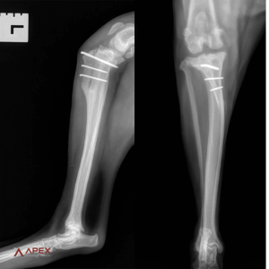
Post-operative lateral and AP radiographs after surgical correction of medial patellar luxation. The pins stabilize the realigned tibial tuberosity, a tibial tuberosity transposition (TTT), one of the techniques used to keep the kneecap seated.
Recovery After MPL Surgery
Recovery varies with the grade of luxation and the procedure performed, but most dogs follow a similar path. The first four weeks center on strict activity restriction, short leash walks only, with no running, jumping, or stairs, to protect the repair while it heals. For procedures that involve a bone cut (such as a tibial tuberosity transposition or a corrective osteotomy), bone healing typically takes around 8 weeks and is confirmed with follow-up X-rays. Controlled physical rehabilitation, gradually reintroduced, helps rebuild strength and range of motion. Most dogs return to normal, comfortable activity within about three months, though your surgeon will tailor the timeline to your dog’s specific case.
When to See a Specialist
Because the right approach depends on the grade, the dog’s age, and the presence of bony deformity, MPL is well suited for evaluation by a board-certified surgeon. Early diagnosis and treatment give the best long-term outcome. Apex Veterinary Surgery’s DACVS surgeons see MPL cases by referral and by direct request at both our Germantown location in the Memphis metro and our Brentwood location in the Nashville metro. Request a consultation.
Frequently Asked Questions
What is medial patellar luxation in dogs?
Medial patellar luxation (MPL) is a condition in which the kneecap (patella) slips inward out of its normal groove at the end of the thigh bone. It’s most common in small and toy breeds and ranges from mild to severe.
What are the grades of patellar luxation?
MPL is graded I to IV. Grade I luxates only with manual pressure and returns on its own; Grade II luxates occasionally; Grade III sits out of the groove most of the time but can be replaced by hand; Grade IV is permanently luxated and cannot be manually repositioned.
Does my dog need surgery for a luxating patella?
Not always. Mild, asymptomatic cases (Grade I and some Grade II) are often managed with weight control, physiotherapy, and activity changes. Grade III and IV, and symptomatic Grade II, usually need surgery for the best outcome.
How successful is MPL surgery in dogs?
Outcomes are generally very good. With appropriate surgery and rehabilitation, most dogs regain comfortable use of the leg, and reported owner satisfaction is commonly over 90%.
What is the recovery time after patellar luxation surgery?
Recovery varies by grade and procedure, but most dogs need roughly 4 to 8 weeks of restricted activity while the repair heals, followed by a gradual return to normal activity guided by controlled rehabilitation. Procedures involving a bone cut are typically rechecked with X-rays to confirm healing.
Which dog breeds are most prone to patellar luxation?
Predisposed breeds include Boston Terriers, Yorkshire Terriers, Pomeranians, Miniature Poodles, and Chihuahuas, though any dog can be affected.
Can a luxating patella lead to other knee problems?
Yes. Over time, repeated luxation can erode cartilage and cause arthritis, and the added strain on the knee can predispose it to a cranial cruciate ligament (CCL) rupture.
Schedule a Consultation
Ready to take the next step? Reach out to book a consult with the top veterinary surgeons in the Midsouth.

Dr. Mac Maxwell is a board-certified veterinary surgeon (DACVS) practicing at Apex Veterinary Surgery’s Germantown office in the Memphis metro area. His training began with a DVM from Mississippi State University College of Veterinary Medicine in 2004, followed by a small animal medicine and surgery internship at Colorado State University Veterinary Teaching Hospital. He returned to MSU for a three-year surgical residency and later served there as an assistant professor of small animal surgery. He became a Diplomate of the American College of Veterinary Surgeons in 2009 and earned a Master’s degree in 2010.
Dr. Maxwell has authored and coauthored book chapters in his field and published peer-reviewed scientific research. He has lectured at continuing education meetings across the region.
Selected publications:
– Duration of use of postoperative thoracostomy tubes in canines and felines without preoperative pneumothorax or pleural effusion, Journal of the American Veterinary Medical Association, 2026. https://doi.org/10.2460/javma.25.09.0603
– Effects of incision closure method on infection prevalence following tibial plateau leveling osteotomy in dogs, The Canadian Veterinary Journal, 2015. https://pmc.ncbi.nlm.nih.gov/articles/PMC4357910/
Medically reviews surgical and orthopedic content on the Apex Veterinary Surgery website.